The Bladder Mechanism
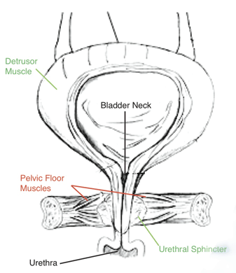
The bladder mechanism consists of the voluntary filling and voiding of the urinary bladder which is controlled by a combined action of the Pelvic Floor muscles (support for the bladder), the Detrusor (muscle surrounding the bladder walls), and the Urethral sphincters (muscles on the Urethra that act as valves for the bladder to be emptied).
During bladder filling, the Detrusor, a smooth muscle, needs to be relaxed (compliant) while the sphincters and urethra need to be contracted. During filling, the Detrusor pressure remains nearly constant.
During voiding, the opposite effect is observed; the Detrusor contracts (its pressure increases) while the sphincters and urethra relax.
The bladder system is complex and our body utilizes a sophisticated feedback control system based on nerves and muscles that process afferent and efferent electrical signals to coordinate various sequences that control Micturition including:
SENSORY INNERVATION
AFFERENT impulses of the bladder wall originate from mechanoreceptors that respond to stretch and free nerve endings in the bladder mucosa that respond to pain and temperature. The impulses travel in the pelvic nerve sand relay on the interneurons in posterior gray horn of the spinal cord from where the secondary neuron travels through the spin thalamic tract to the Pontine micturition center PMC. Afferent impulses originating at the trigon and urethra relay to the cerebral cortex.
MOTOR INNERVATIONS OF THE BLADDER
SOMATIC MOTOR innervation to the external urethral sphincter and pelvic floor muscles is provided by the ONUFs nucleus, which is located in the anterolateral horn of the sacral spinal cord (S2, S3, S4). The fibers travel through the Pudental nerve. The Parasympathetic and Sympathetic Nervous Systems work synergistically (opposite effects) as they control the bladder and urethra.
-
The Preganglionic Parasympathetic nerves from S2, S3 and S4 travel in the pelvic nerve and synapse in the cholinergic ganglia in the pelvic plexus. They also provide the major motor innervation to the Detrusor muscle (wall of the bladder). The Parasympathetic nervous system (PNS), through its effect on cholinergic receptors in the bladder and urethra, excite the DETRUSOR and inhibit URETHRAL smooth muscles.
-
The Preganglionic Sympathetic nerves from T10 to T12 com are the conduit for postganglionic neurons to travel in the hypo gastric nerve and synapse in the adrenergic ganglia in the pelvic plexus. The Sympathetic nervous system (SNS) via its effect on beta adrenergic receptors inhibits DETRUSOR and stimulates urethral smooth muscle via Alfa adrenergic receptors.
CENTRAL CONTROL
Neural pathways play a critical role in storage and voidance of the bladder. The Parietal lobes and Thalamus receive and coordinate Detrusor afferent stimuli, the Frontal lobes and Basal ganglia provide modulation with inhibitory signals, thus providing tonic inhibitory system in the brain that suppress Parasympathetic excitatory outflow to the bladder. Damage to central inhibitory pathways can unmask primitive voiding reflex and trigger bladder over reactivity.
Pontine micturition center (PMC) is located in gray area of the pons, and is the area of integration and control of afferent and efferent impulses from the urinary bladder. The Pontine continence center (PCC) is located in reticular formation of the pons. Its impulses provide a tonic excitation to the motor neurons of ONUF s nucleus, thus the pelvic floor muscles and striated urethral sphincter maintain a baseline tone.
When PMC is stimulated it activates sacral inhibitory interneurons, which in turn, inhibit motor neurons to the external urethral sphincter and pelvic floor muscles. Urethral pressure is thus decreased and pelvic floor muscles relaxed. By simultaneously stimulating Parasympathetic innervation of the detrusor, PMC causes increase in bladder pressure and allows micturition to take place.
What this means for bladder function
Filling and voiding involves anatomic and neurologic mechanisms described below. Alternations in any of these components may result in dysfunctional filling / voiding and /or urinary incontinence:
FILLING AND STORAGE OF URINE
The Detrusor and bladder constitute an extremely compliant organ (IntraVesical pressure increases minimally during a bladder filling).
In response to afferent neurons excitation during advanced filling, the SNS inhibits Detrusor contractions by relaxing the smooth muscle through beta Adrenergic receptors. At the same time Sympathetic inhibition of Parasympathetic ganglionic transmission in the Pelvic Plexus and activation of the Urethral smooth muscles through Alfa adrenergic receptor take place facilitating the storage of urine.
Bladder stretch receptors communicate with PCC, which also increases SNS efferent activity, as well as activates ONUF s nucleus motor neurons to amplify tone of the striated muscles in the urethra and pelvic floor.
MICTURITION (Voiding)
When the bladder capacity is reached, the bladder afferent nerve signals activate PMC which, in turn stimulates PNS efferent signals. This results in relaxation of external Urethral sphincters and decrease in Pelvic Floor muscles pressure during the contraction of the Detrusor (VOIDING).